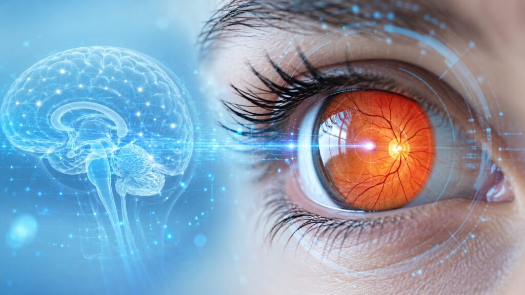
People at risk of dementia can be identified by their eyes
Alzheimer’s disease develops over decades, but symptoms only appear when the brain has already sustained irreversible damage. Recently, scientists discovered that the risk of developing dementia can be predicted long before the first memory problems appear. All it takes is analyzing ordinary retinal fundus images using artificial intelligence.
How the Retina Reflects the State of the Brain
The retina is a light-sensitive tissue on the back wall of the eye that contains several layers of neurons and a dense network of tiny blood vessels. In fact, it is a direct extension of the nervous system.
Therefore, any changes in the retinal blood vessels reflect hidden diseases within our body. Already today, fundus photographs are regularly taken for patients with diabetes and glaucoma. However, scientists from Florida have proven that these images contain far more useful information.
The retina works as a biological sensor built into our body. If a pathology begins to develop in the brain, its first traces can be noticed simply by looking into a person’s eyes.
Searching for Signs of Alzheimer’s Disease with Neural Networks
To find the connection between vision and the brain, a team of scientists used machine learning. Neural networks analyzed nearly 63,000 retinal photographs belonging to more than 40,000 participants of the large-scale medical project UK Biobank.
The artificial intelligence was trained to predict 12 key factors associated with an increased likelihood of developing dementia. Among them:
- age and body mass index;
- sleep quality and blood pressure levels;
- tendency toward depression;
- harmful habits such as smoking and alcohol.
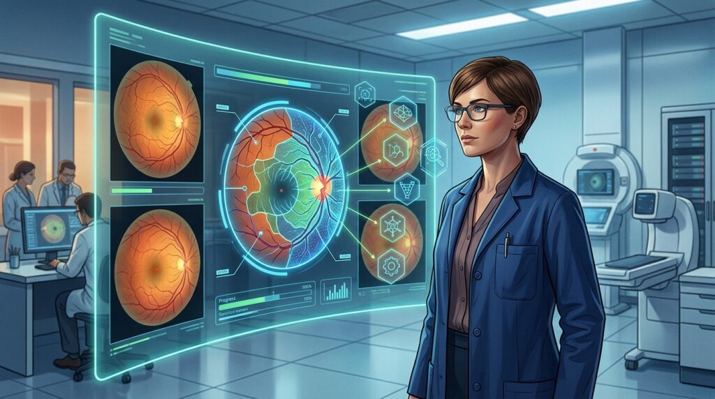
Artificial intelligence can find patterns in eye images that are invisible to a doctor
As a result of deep learning, the algorithm learned to detect the subtlest markers of the upcoming disease, combining risk factors into a unified picture.
Hidden Changes in Eye Blood Vessels Associated with Dementia Risk
In patients who were subsequently diagnosed with Alzheimer’s disease, artificial intelligence identified specific features of the fundus. These changes occur years before a person begins to forget names or lose spatial orientation.
The main indicator was narrowing of small arteries and arterioles in the retina. In addition, the algorithm detected decreased density of the vascular network, stiffness of vessel walls, and thinning of the optic nerve.
Such symptoms indicate disruption of the circulatory system that feeds nervous tissue. Although at this stage this is only an association rather than a precise diagnosis, weakening of the eye’s blood vessels clearly coincides with long-term deterioration of brain function.
How Retinal Analysis Will Change Early Alzheimer’s Diagnosis
It is important to understand that a retinal photograph cannot provide an Alzheimer’s disease diagnosis ten years before symptoms appear. However, this method excels at another task — it identifies people in the high-risk group.
Currently, the main problem in the fight against dementia is that treatment often begins too late. If risk can be determined during a routine visit to an ophthalmologist, patients will gain precious time for prevention.
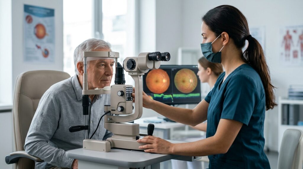
In the future, a routine eye exam could become the first step in dementia prevention
By becoming aware of their predisposition to Alzheimer’s disease, a person can change their lifestyle: adjust their diet, normalize sleep, and lower blood pressure. It is quite possible that in the coming years, retinal images will become a universal tool for monitoring brain health.
Alzheimer’s disease leaves a kind of “breadcrumb trail” as it develops silently. Studying the retina helps scientists follow this trail to better understand the nature of dementia and learn to stop it before memory is affected.