Scientists proved: the human heart is capable of recovering after a heart attack
If you’ve ever heard a doctor say that heart muscle doesn’t recover after a heart attack — that’s not entirely true. The human heart can grow new muscle cells after a heart attack — and this has recently been proven in humans for the first time. However, it’s too early to celebrate: natural regeneration isn’t sufficient to fully heal the heart, but the very fact opens doors for new methods of treating heart failure and attempts to teach the heart to heal itself.
Why the Heart Was Considered Incapable of Recovery
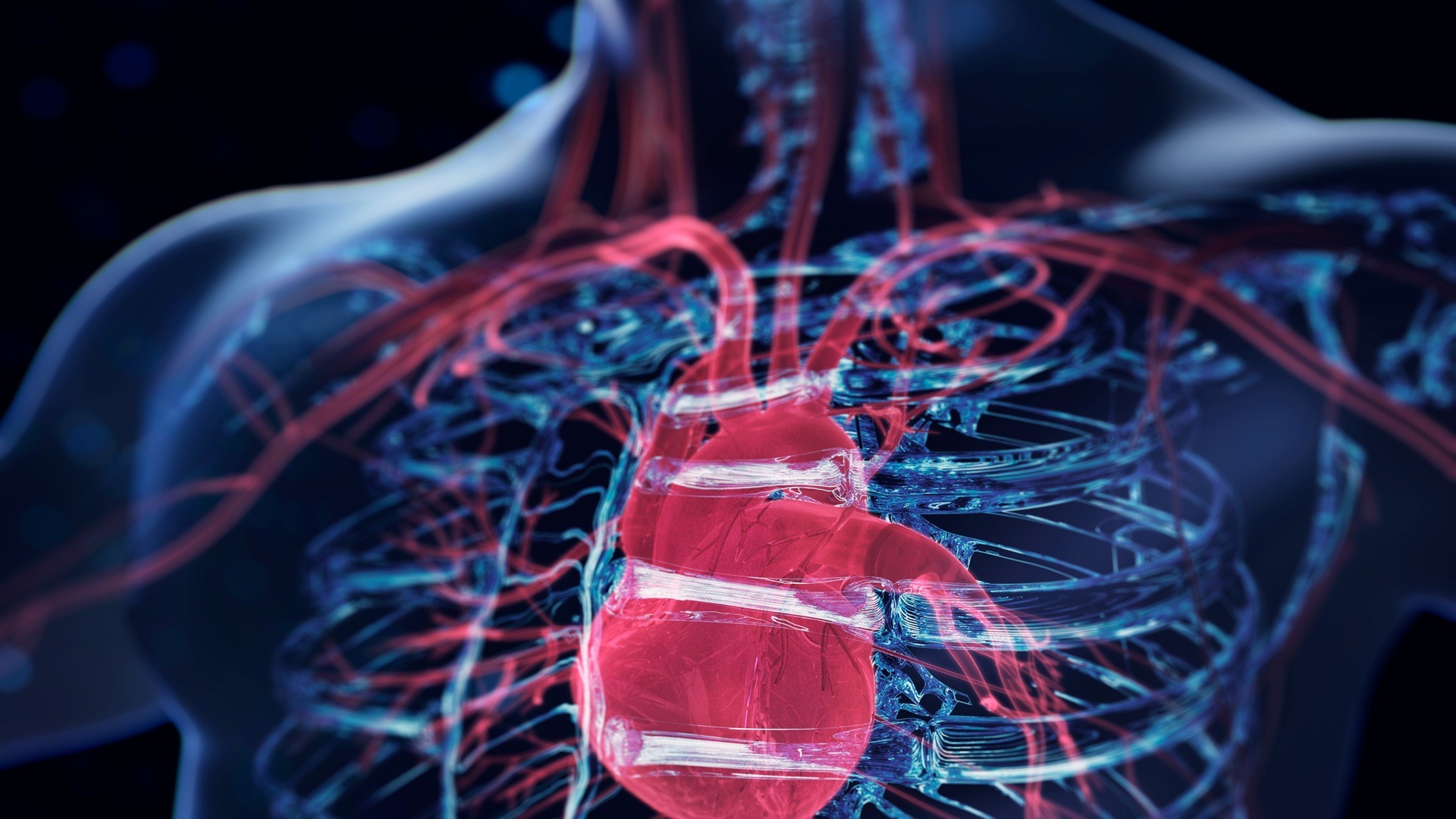
Heart muscle cells — cardiomyocytes — are not structured like skin or liver cells. Skin cells divide constantly, and the liver can regrow a lost portion. But cardiomyocytes behave differently: they actively divide only during fetal development, and shortly after birth they almost completely stop dividing. This is precisely why medical textbooks stated for decades: the number of heart cells you’re born with is roughly the same number you’ll die with (unless some are destroyed by a heart attack).
A myocardial infarction can destroy up to a third of all cardiomyocytes. In place of the dead cells, a scar forms — similar to a scar on the skin, except a scar made of connective tissue cannot contract, meaning the heart begins to pump blood less effectively. Over time, this can lead to heart failure, the only radical treatment for which remains transplantation. But donor organs are chronically in short supply, which is why medicine is simultaneously developing artificial hearts as well.
Meanwhile, experiments on mice showed a quarter century ago that after a heart attack, rodent cardiomyocytes begin dividing more actively. But confirming the same thing in humans had proved impossible — it was too difficult to obtain and study living human heart tissue.
How Scientists Proved Heart Regeneration After a Heart Attack
The study was conducted by a team from the University of Sydney, the Baird Institute, and Royal Prince Alfred Hospital. Their work was published in the journal Circulation Research.
The starting point was a unique case. Professor Sean Lal, head of the Sydney Heart Bank (a repository of cryopreserved heart tissue), discovered a rare specimen in the collection — the heart of a 48-year-old man who had suffered a massive heart attack due to complete blockage of the left anterior descending artery. The patient was on life support for five days and was declared dead by neurological criteria, and his heart, unsuitable for transplantation, was donated for research before circulatory arrest.
Such a “premoral” (ante-mortem) heart with an infarct zone is exceptionally rare. As the authors themselves note, the probability of obtaining a similar specimen again is extremely low. Therefore, the team developed an additional method: they began collecting live tissue biopsies from patients undergoing coronary artery bypass surgery 7–10 days after a heart attack.
How Heart Cells Divide After a Heart Attack
The researchers used a full arsenal of methods — immunohistochemistry, RNA sequencing, proteomics, metabolomics, and single-nucleus sequencing. All of this allowed them not just to see dividing cells, but to study in detail which genes and proteins are activated during regeneration.
The result was unequivocal: adult human cardiomyocytes increase mitosis (cell division) in response to ischemia — the oxygen deprivation caused by a heart attack. In samples from the zone adjacent to the infarct, about 7–8% of cardiomyocytes showed signs of division, and in the unique “premoral” heart, this figure reached up to 11%.
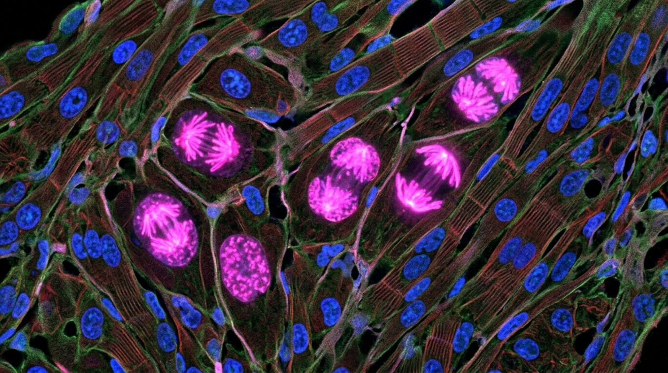
Fluorescence microscopy of heart tissue: dividing cardiomyocytes highlighted in pink
It’s important to understand the scale: for full heart recovery, according to Professor Lal’s estimate, 25–50% of cells would need to be dividing. The actual 7–11% is significantly less, but it radically changes the very understanding of what the human heart is capable of. Interestingly, similar mechanisms of tissue recovery after damage are being found in other organs as well, suggesting a broader potential for regenerative medicine.
Why Oxygen Deprivation Triggers Heart Recovery
One of the most interesting hypotheses of the study is related to the mechanism that triggers regeneration. Professor Lal suggests that the trigger is hypoxia itself — the very oxygen deprivation that kills some cells during a heart attack.
The logic is as follows: during fetal development, the fetus is in a low-oxygen environment, and it is precisely during this period that cardiomyocytes actively divide. After birth, when the lungs begin working and oxygen levels rise sharply, heart cell division almost completely stops. It’s possible that during a heart attack, oxygen deprivation “awakens” the same program in heart cells that operated in the embryo. Analysis showed that genes active in fetal cardiomyocytes were “switched on” only in the zone adjacent to the damage.
This is still a hypothesis, not a proven fact. But if confirmed, it could suggest how to enhance regeneration — for example, with drugs that mimic the effect of controlled hypoxia.
How the Discovery Will Change Heart Failure Treatment
Cardiovascular diseases remain the leading cause of death worldwide. For example, in Russia, chronic heart failure affects 8.2% of the population — roughly 12 million people — while only 522 heart transplants were performed in 2025. The gap between need and capacity is enormous.
The Australian team’s discovery doesn’t solve this problem instantly, but it changes the paradigm itself. Previously, the conversation was framed as “the damage is irreversible.” Now — “the damage is partially reversible, and this ability can potentially be enhanced.”
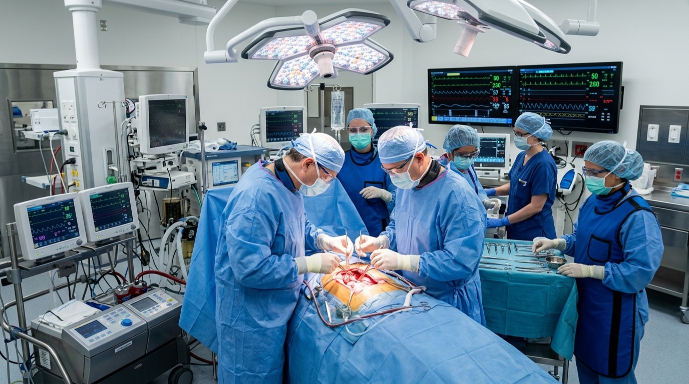
Heart surgery in a modern clinic
The researchers have already identified several proteins in human tissue samples previously linked to heart regeneration in mice. This means that biological recovery pathways may be shared across different mammalian species, and findings from rodent experiments won’t have to start from scratch. Previously, similar hopes were tied to stem cells for the heart, but it turned out that such processes are more complex than simply replacing dead tissue. Additionally, the developed method of live tissue biopsy gives scientists a working model for testing future regenerative drugs directly on human cells.
Dr. Robert Hume himself, the study’s first author, emphasizes: natural regeneration is still too weak to prevent the severe consequences of a heart attack. But the goal is to develop a therapy that will enhance the heart’s innate ability to self-repair and, in the long term, allow reversing heart failure without transplantation.