A new method of computed tomography allows for more detailed study of Egyptian mummies
At Budapest’s Semmelweis University, several Egyptian mummies that had spent decades in museum collections were scanned using a CT scanner with a photon-counting detector. This is a next-generation device typically used for examining living patients. The results have already overturned some previous assumptions about what exactly the collection contains. One find, previously thought to be a head and later reclassified as a bird mummy, turned out to be a human foot.
What Was Found Inside Egyptian Mummies in Budapest
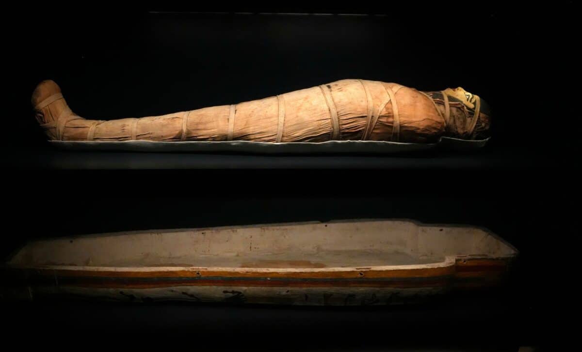
The specimens for the study were provided by the Semmelweis Museum of the History of Medicine, part of the Hungarian National Museum. The collection includes two mummified heads, two left lower limbs, a bundle containing a mummified foot, and a hand. These are all body parts rather than complete mummies — the remains presumably once belonged to whole mummies, but when and why they were separated remains unknown.
Previously, researchers attempted to date the six specimens using radiocarbon methods, but only three of the six yielded usable results. According to these data, the oldest remains date to between 401 and 259 BCE, making them over 2,300 years old.
The very first scanning results yielded concrete findings. Images of one of the lower limbs indicate osteoporosis — a disease in which bones lose density and become brittle. Previous studies using conventional CT scanning had not been able to make such a diagnosis. The second lower limb, as it turned out, belonged to a young person, which also could not be determined before.
How the CT Scanner Studies Egyptian Mummies
To understand why the new scanner sees more, it’s worth examining how it differs from a conventional one. In a classic CT machine, X-ray radiation first hits a scintillator — a material that converts X-rays into visible light. The light is then converted into an electrical signal. The problem is that this approach registers the total energy of many photons at once along with electronic noise, and fine details are lost — roughly like trying to hear individual voices in a noisy crowd while recording only the overall sound level.
A photon-counting detector works on a fundamentally different principle: it registers each individual X-ray photon and records its energy. A semiconductor crystal directly converts the photon into an electrical signal, bypassing the visible light stage. Since the signal is not subject to afterglow and decay, the technology allows clear separation of useful information from electronic noise.
In practice, this provides several advantages simultaneously: higher spatial resolution, better soft tissue contrast, and a lower radiation dose. In the case of the Budapest scanner, the radiation dose is nearly half that of conventional CT, and details as small as a few millimeters become visible. For mummies, where it’s necessary to distinguish bone, resin, linen layers, and air pockets, this is critically important.
The first photon-counting CT scanner for clinical use was approved by the FDA in September 2021. Semmelweis University has already put three such devices into operation — and uses them not only for patients but also for scientific research.
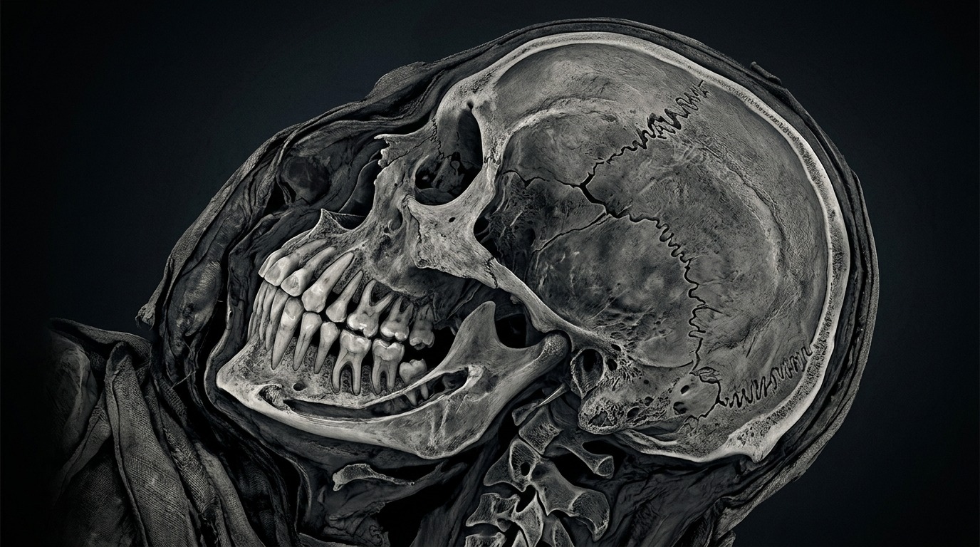
Detailed CT scan of a mummified head with visible teeth and cranial sutures
Bird Mummy Turned Out to Be a Human Foot
One of the most unexpected findings involves a bundle of bandages that the museum received with no visual clues about its contents. When the find entered the collection, it was first described as a human head, and later reclassified as a bird mummy. However, a previous CT scan had already shown that inside was an adult human foot.
The new generation of CT scanner went further. Researchers are now studying the textile fragments of the wrapping to understand the mummification technique, the person’s age, and possible diseases. The scans clearly show different layers of bandages and their structural features. This is important because bandaging techniques changed over time and varied depending on the social status of the deceased.
The story of the “foot that was mistaken for a head” illustrates well why museum collections should be revisited with new tools. When an object has been described by eye for centuries, errors accumulate — and only non-invasive imaging allows them to be corrected without damaging the artifact.
Teeth, Bones, and 3D Facial Reconstruction of Ancient Egyptians
The high resolution of the new scans allows detailed examination of the teeth and cranial sutures of two mummified heads. Cranial sutures are the junctions where skull bones meet, which gradually fuse with age. The more precisely their contours can be seen, the more accurately the person’s age at death can be estimated.
This data can help refine age estimates and serve as a basis for creating accurate 3D models, including possible facial reconstructions. In effect, scientists are getting closer to looking into the face of a person who lived in Egypt during the Ptolemaic period.
A separate challenge is a mummified hand whose owner has not yet been identified. Based on the size and shape of the bones, researchers are trying to determine whether this was the hand of a child or an adult. With previous equipment, distinguishing between these options was not possible.

Researchers analyze a three-dimensional bone model on a monitor
Why Museum Mummies Need to Be Re-Examined
Everything described above was obtained from objects that have been stored in the museum collection since its founding and have been repeatedly examined in recent years using various methods, including conventional computed tomography, but technological limitations prevented obtaining a sufficiently detailed picture.
This is an important lesson not only for Egyptology. Thousands of museums around the world hold remains that were described and catalogued decades or even centuries ago. Modern imaging can reveal hidden details without cutting or unwrapping anything, and for museums, non-invasive scanning is a way to protect fragile remains while simultaneously verifying old descriptions.
Detailed analysis of the obtained images is still ongoing, and a number of preliminary conclusions remain precisely that — preliminary. For definitive diagnoses, researchers need to compare the scans with skeletal anatomy standards, textile data, and previous collection records. So there is still plenty of work ahead.
But the main takeaway is already clear: technology created for diagnosing diseases in living people has proven to be a powerful tool for studying people who lived more than two thousand years ago. The trend of using cutting-edge medical imaging to study ancient remains is gaining momentum — and the Budapest project shows that even well-studied collections can hold surprises when viewed with fresh eyes and the right equipment.